
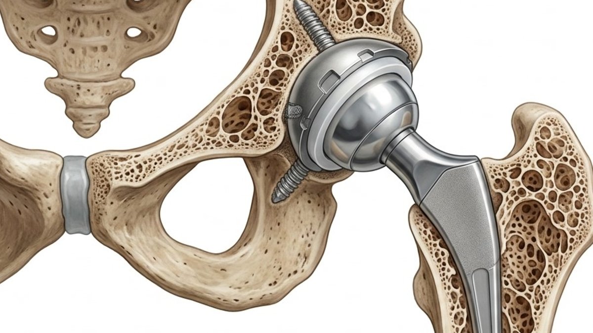
New research from the International Osteoporosis Foundation (IOF) identifies brittle bone as a key factor behind implant failures in hip replacements and spinal fusions. The position statement, published in Osteoporosis International on 18 June 2026, argues that osteoporosis and implant failure are interconnected, urging surgeons to screen and treat bone health before and after these procedures.
A Shared Underlying Problem
The IOF’s Fracture Working Group defines “implant fixation failure” as loss of mechanical integrity in the bone-implant unit over time. This includes periprosthetic fractures, aseptic loosening, cage subsidence, and pseudarthrosis. These complications, the authors argue, stem from bone that cannot withstand the altered loading of metal implants.
Between 60 and 80 percent of patients undergoing knee replacements have osteopenia or osteoporosis, yet nearly 75 percent remain undiagnosed pre-operatively. Periprosthetic femoral fractures carry an 11 to 18 percent one-year mortality rate, comparable to native hip fractures. In spinal fusion, a systematic review of 71 studies involving over 12,000 patients found low bone mineral density (BMD) linked to cage subsidence, screw loosening, and proximal junctional kyphosis from fracture.
Screening and Treatment Recommendations
Patients aged 65 and over, those with prior fragility fractures, or individuals undergoing high-demand procedures like revision arthroplasty or long-segment spinal fusion should have bone health assessed pre-operatively. The working group recommends integrating bone screening into routine surgical pathways, alongside standard cardiac and anaesthetic evaluations. Surgery should not be delayed for this assessment, a strong recommendation based on registry and observational data.
Related Post: Genetic Screening Could Reveal Early Cancer Risk in Newborns
Bisphosphonates are advised post-operatively for arthroplasty patients with osteoporosis or high-risk bone phenotypes, with denosumab as an alternative where bisphosphonates are contraindicated. Discontinuing denosumab without transitioning to another antiresorptive risks rebound bone turnover and vertebral fractures. For spinal fusion, teriparatide—typically started at least a month before surgery and continued for six to twelve months—shows the strongest evidence for higher fusion rates and reduced pedicle screw loosening. Romosozumab, while supported by retrospective and preclinical data, is time-limited and should complement broader osteoporosis strategies.
Diagnostic Challenges and Emerging Tools
Current diagnostic methods often overlook mechanical bone quality around implants. Standard dual-energy X-ray absorptiometry (DXA) scanning, while the reference standard, fails to capture regional bone loss. The paper notes a 15 percent reduction in distal femoral bone density within six months of knee replacement, concentrated at the implant interface. However, methodology heterogeneity in existing studies complicates precise quantification.
Alternative approaches show promise. Opportunistic CT scans taken during surgical planning can calculate Hounsfield units, offering region-specific bone quality data. Gruen-zone specific DXA analysis after hip replacements also provides targeted insights. These methods are not yet widely available but may better reflect implant-related bone changes than global BMD measurements. Radiographic and biomechanical surrogates currently dominate research outcomes, limiting conclusions about long-term clinical impact.
Evidence Gaps and Future Directions
The authors emphasize the need for outcome-driven trials to validate treatment strategies. Most studies rely on surrogate endpoints like bone density changes or screw pull-out strength, rather than revision rates or fracture prevention. Systematic peri-operative bone health optimization may enhance construct longevity, but definitive evidence remains lacking. Collaboration between orthopaedic surgeons and osteoporosis specialists is essential to advance this field.


Leave a Reply